2007-10-25
Which areas of the brain say “ouch”? An fMRI study exploring the neural basis of pain.
Publication
Publication
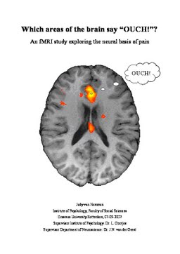
The aim of this study was to a) determine which brain areas are involved with the processing of painfully hot stimulation and b) investigate whether individualized and standardized painfully hot stimuli lead to the same activation patterns. Nineteen participants underwent functional magnetic resonance imaging (fMRI) scanning while receiving neutral (32°C), warm (37°C) and painfully hot stimuli on the hand. Half of the painfully hot stimuli were standardized (46°C) and the other half were individualized, using the pain threshold temperature as stimulation temperature (46-48°C). Significant increases of activation during individualized painfully hot stimulation compared to baseline were observed in the insula, anterior cingulate cortex, amygdala, basal ganglia, orbital part of the frontal gyrus, rolandic operculum, superior temporal pole and the superior temporal lobe. These results show great overlap with previous research using fMRI or positron emission tomography (PET). When comparing standardized and individualized painfully hot stimulation, no significant differences in activation were found. The results suggest that the used protocol is a good tool to investigate the neural basis of pain.
| Additional Metadata | |
|---|---|
| , , | |
| Gootjes, L., Strien, J.W. van | |
| hdl.handle.net/2105/4224 | |
| Psychology | |
| Organisation | Erasmus School of Social and Behavioural Sciences |
|
Hemmen, J. van. (2007, October 25). Which areas of the brain say “ouch”? An fMRI study exploring the neural basis of pain.. Psychology. Retrieved from http://hdl.handle.net/2105/4224 |
|